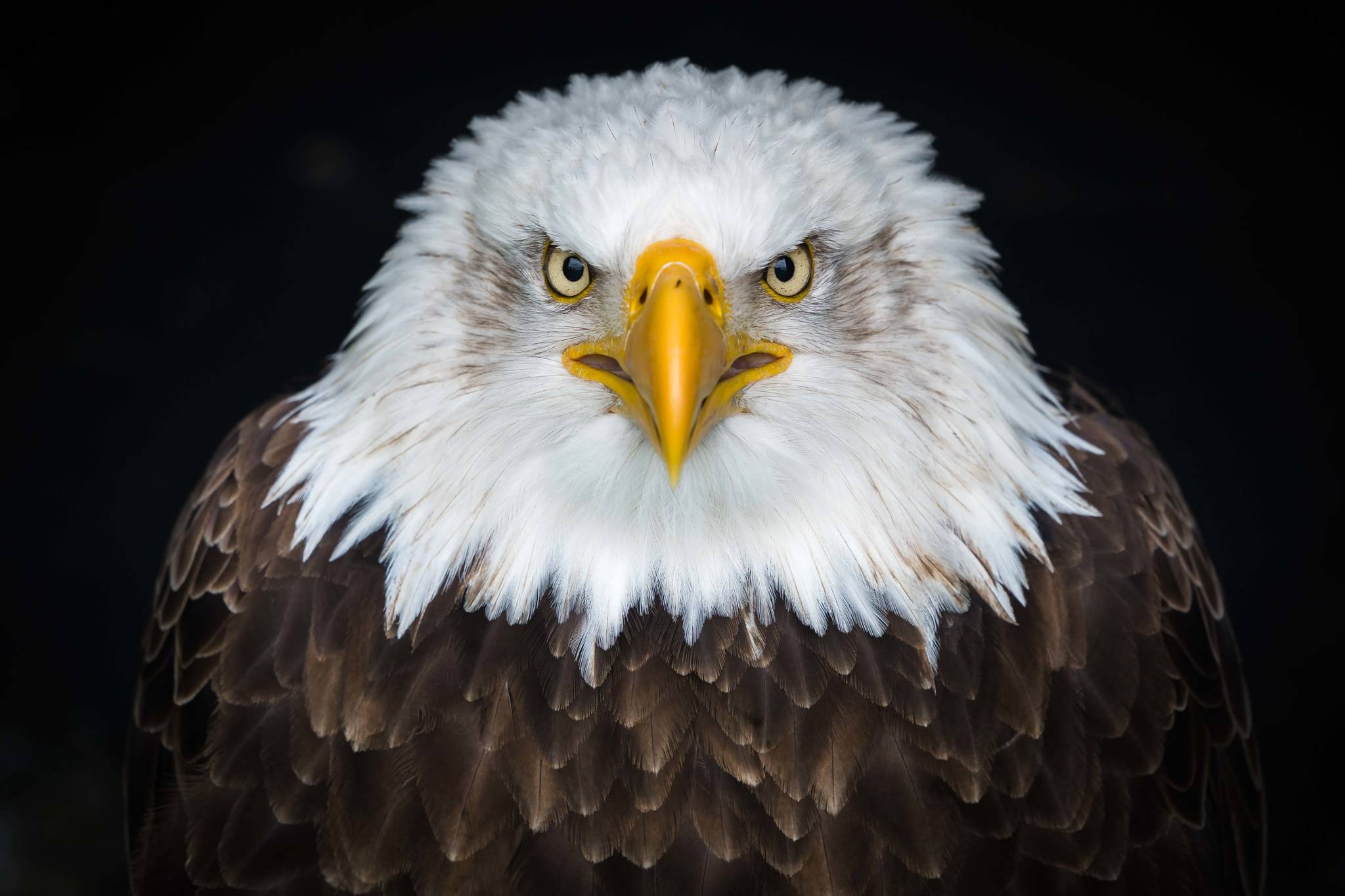
We say that a sharp-eyed person has “the eyes of an eagle.” Eagles are reported to be able to spot a rabbit from a distance of two miles. Eagles, hawks and many other birds are well-known for their visual acuity. Acuity is defined as the ability to discriminate two points as separate entities from a distance; more particularly, it refers to the ability to discern shapes.
For that ability to be brought into play, however, the eye must be able to focus. In humans and other mammals, visual focus is achieved by changing the shape of the lens, bending the incoming light rays so they hit the retina. But in most birds, both the lens and the cornea can change shape. That potentially gives such birds an especially good range of focus (diving birds are an exception — the cornea has the same refractive index as water, so changing its shape would have no effect on focus, and only the lens is used in these birds).
How does visual acuity work?
Visual acuity depends on the function of three interconnected but distinct levels. The first level is the retina, where millions of photosensitive cells line the back of the eye. In the second level, those cells are connected by short nerve fibers to nerve cells that are sometimes clustered together in ganglia, where they are often connected to each other. And in the third level, those ganglia are connected, via more nerve fibers, to the optic centers of the brain via the optic nerve, essentially a cable of millions of long nerve fibers. Here is a brief rundown about those three functional levels (structurally, the peculiar arrangements in vertebrate eyes are more complex and better omitted for present purposes).
Level 1. There are two kinds of photosensitive cells in the retina. Rods (so-called for their shape) are very sensitive and work well in dim light. The eyes of nocturnal birds and many mammals have mostly rods. Cones (spindle-shaped cells with a cone-shaped point where the photosensitive pigments are located) work best in relatively bright light and provide sharp resolution and color vision. Birds typically have four kinds of cone cells, sensitive to red, green, blue and ultraviolet (UV) light, while mammals have only three kinds (no UV). Human eyes have millions of cone cells; avian eyes can have more than that, the numbers depending on both the size of the eye and the density of cones.
Cone cells are densest in an area of the retina called the fovea, which shows up as a small dip in the retinal surface. The eyes of humans and other primates have one fovea, as do most birds. Some mammals and birds (e.g., chickens and quail) have no fovea at all. Terns, swallows, swifts, hummingbirds, hawks, eagles and kingfishers are all reported to have two foveae in each eye. One fovea deals with binocular vision aimed at a target, as when a hawk, kingfisher, or a swallow is pursuing its prey; the other deals with sideways monocular vision for spotting prey at a distance.
Level 2. The number of cone cells connected to a ganglion varies greatly. In the fovea, there may be just one cone per ganglion, but in the rest of the retina, the ratio might be a hundred cones to one ganglion (for example). Clearly, the more cones connect to a single ganglion, the less precise the visual resolution is likely to be. However, within a ganglion, the nerve cells sometimes inhibit each other or perhaps interact in other ways, so the cone/ganglion ratio is not the only factor.
Level 3. The long fibers of the ganglia transmit signals to the brain, which obviously must be wired to somehow interpret and make use of the incoming information. On the way to the interpretive centers of the brain, the optic nerves of vertebrates partially cross, such that some of the nerve signals from the left eye are delivered to the right side of the brain, and some of the signals from the right eye are delivered to the left side of the brain.
The proportion of nerves that cross varies among species. This may be related to, among other things, the ability to coordinate movements of the limbs. Many birds are reported to be able to sleep on one side of the brain while keeping awake and alert (with the eye frequently open) on the other side. Some individuals exhibit a preference for one side or the other. Siamese cats, noted for their usually crossed eyes, are said to have a defect in the optic-nerve crossover that makes the eyes try to compensate for the faulty signals.
Sharpness of vision may be assisted by other adaptations of the avian (but not the mammalian) eye. For instance, many day-active birds have oil droplets in the cones of the eye. The droplets are red, yellow or clear; different kinds of birds and even birds in different populations of the same species may have different oil droplets. The droplets are light filters that can change the light rays that actually reach the retina. For example, they can narrow the range of wavelengths registered by the cones, increasing the color contrast. Sharpening the color contrast might improve discrimination of shapes in some circumstances.
Visual acuity is a matter of spatial resolution. But many birds also have excellent temporal resolution; they can process temporal changes in light very quickly. For comparison, humans cannot detect separate light pulses that flicker faster than about 50 times per second — faster flickers are just a blur to us. But birds can process fast flickers and rapid movements more quickly than we can. This may allow them to register wing beats of a prey insect, for example, or detect the location of twigs and branches as they fly through the tree canopy.
• Mary F. Willson is a retired professor of ecology. Her essays can be found online at www.onthetrailsjuneau.wordpress.com

